
소득공제
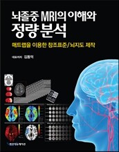
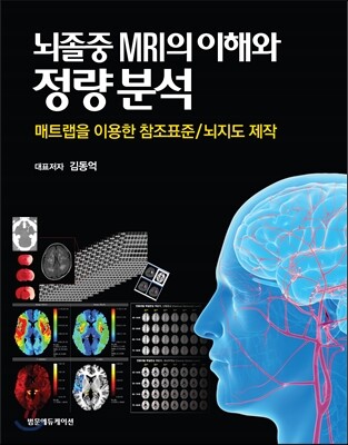
뇌졸중 MRI의 이해와 정량분석
매트랩을 이용한 참조표준/뇌지도 제작
- 베스트
- 의약학/간호 계열 top100 1주
- 교재철(3월,9월) 소량 입고도서로 배송지연 혹은 주문이 취소될 수 있습니다.
이미 소장하고 있다면 판매해 보세요.
- 국내배송만 가능
- 수급불안정으로 인해 품절/지연 가능
- 문화비소득공제 가능
책소개
목차
|
CHAPTER 1. 들어가기 1
Introduction 참고문헌 4 CHAPTER 02 자기공명영상의 원리 5 Principles of Magnetic Resonance Imaging 1. 자기공명영상(MRI)이란? 5 자기공명영상(MRI) 기본이해 5 스핀에코 시퀀스 (spin-echo sequence) 8 기울기에코 영상 (gradient-echo imaging; GRE) 10 FLAIR (fluid-attenuated inversion recovery) MRI 11 조영증강영상(contrast enhanced imaging) 11 확산강조영상 (diffusion-weighted MRI; DWI) 14 2. 뇌경색 MR 영상 및 뇌병변 분할/정합 16 뇌경색 코어(Infarct Core) 분할 16 FLAIR MR 영상 18 T2강조영상 (T2-weighted MRI) 18 경사에코 영상 18 관류강조영상 정량화 18 3. 뇌 템플릿 정합 19 참고문헌 21 CHAPTER 03 뇌졸중 뇌자기공명영상 판독 23 Magnetic Resonance Imaging in Stroke: Diagnosis & Therapeutic Implications 1. 확산강조영상 24 뇌경색의 위치, 분포, 체적 평가 25 추적 검사에서 새로운 뇌졸중 관련 병변의 발생 30 확산강조영상에서 보이는 고신호강도 병변과 액체감쇠역전회복영상에서 보이는 병변의 일치 여부 33 확산강조영상에서 확산이 제한되어 뇌경색과 구분이 필요한 경우 34 2. 액체감쇠역전회복영상 34 뇌경색과 혈관주위공간의 구분 36 소량의 거미막하출혈 진단 38 고신호강도혈관소견 38 혈액뇌장벽(blood-brain barrier) 손상 여부 판단 40 T2 강조영상 40 T1강조영상 41 위상에코영상 44 관류 영상 44 혈관영상 49 참고문헌 51 색인 52 CHAPTER 04. 한국 뇌경색 환자의 뇌백질변성과 뇌졸중 위험인자 및 뇌경색 예후와의 연관성 53 The Links between White Matter Hyperintensities and Stroke Risk Factors or Post-stroke Outcomes in Korean Patients with Cerebral Infarction 1. 뇌백질변성 (white matter hyperintensities of presumed vascular origin) 53 2. 뇌백질변성의 기전 55 3. 뇌백질변성의 위험인자 55 4. 뇌백질변성과 초급성기 뇌경색 치료 58 5. 뇌백질변성과 뇌졸중의 예후 60 6. 뇌백질변성과 뇌졸중의 발생 및 재발 64 참고문헌 65 CHAPTER 05. T2*경사에코영상 및 자화강조 영상을 이용한 미세출혈 진단 69 Detection of Cerebral Microbleeds using T2* Gradient-echo Imaging and Susceptibilityweighted Imaging 1. T2* 경사에코영상 (T2* gradient echo imaging, GRE) 69 GRE 영상의 원리 69 시퀀스의 종류: gradient echo (GRE) 혹은 spin echo (SE) 70 에코 시간의 효과 71 자기장 강도의 효과 71 공간 해상도 72 2. 자화강조영상 (Susceptibility-weighted imaging) 72 3. 미세출혈(Cerebral microbleeds) 73 미세출혈의 진단 73 미세출혈의 병리와 분포 74 미세출혈의 역학과 위험인자 75 미세출혈의 임상적 의의 76 4. 맺음말 78 참고문헌 78 CHAPTER 06 Grading and Interpretation of White Matter Hyperintensities Using Statistical Maps 83 ABSTRACT 84 Introduction 85 Materials and Methods 87 Study population 87 Clinical database 89 Brain MRI and quantitative image registration 89 Statistical anatomic maps: WMH frequency-volume maps 96 Statistical analysis 96 Results 99 Patients and WMHs 99 Frequency-volume maps 99 Associations between WMH volume and risk factors 99 Analyses after two-by-two stratification by hypertension and age (#69 vs.$ 70) 105 Discussion 108 Conclusion 115 Acknowledgments 115 Sources of Funding 115 Disclosures 115 References 116 CHAPTER 07 Stroke Outcomes Are Worse With Larger Leukoaraiosis Volumes 121 ABSTRACT 122 Introduction 123 Materials and methods 124 Participants 124 Clinical data collection 124 Definition of early neurological deterioration and late recurrence of stroke 125 Magnetic resonance image registration and analysis 126 Data analysis and statistics 127 Results 129 Baseline characteristics and univariate analyses 129 Association between WMH quintiles and 3-month modified Rankin Scale for the entire study population 130 Association between WMH quintiles and 3-month modified Rankin Scale for each stroke subtype 130 Association between WMH volume and the initial neurological severity at admission 134 Association between WMH quintiles and the incidence of early neurological deterioration 137 Association between WMH quintiles and early or late stroke recurrence 140 WMH quintiles and functional recovery from discharge to 3-month 140 Early neurological deterioration-adjusted 3-month modified Rankin Scale score 140 Discussion 142 Conclusion 148 Funding 148 References 149 CHAPTER 08 매트랩을 이용한 뇌 MR영상 정량분석 및 매핑 153 Matlab-based Quantitative Brain MRI Analysis and Mapping c:\data\bmp2nii\m_file\mk3DNIfTI_with_2DBMPs_316S4one_folder_final.m 154 c:\data\templates\24_slices_nii_templates\mk6mm_thick_24_slicesTemplate.m 160 c:\data\make24s_nii\m_file\mk3D24slices_from_316S.m 162 c:\data\mkFreqMap\m_file\mkFreqMapWithNIfTI.m 166 c:\data\mkFreqMap\m_file\overlayMap.m 168 c:\data\quantification\count_lesion_voxels_on_24S_nii\ count_lesion_voxels_on_24S_nii.m 172 c:\data\quantification\count_lesion_voxels_on_bmp\ count_lesion_voxels_on_BMP_file.m 174 c:\data\quantification\calculation_of_vol_percentage\calVolPct.m 176 c:\data\quantification\calculation_of_vol_percentage\calTotalVolume.m 181 c:\data\mkFreqVolMap\mkFreqVolMap.m 183 c:\data\mkFreqMapWithBMP\m_file\mkFreqMapWithBMP.m 190 c:\data\mkFreqMapWithBMP\m_file\overlayMap4BMP.m 191 c:\data\quantification\vox\sl_sel.m 193 참고문헌 195 APPENDIX I. 만성 뇌허혈 (대뇌 백질변성) 참조표준 뇌지도 Atlas 197 APPENDIX II 뇌졸중 뇌MR영상 참조표준 Tables 217 1. FLAIR MRI: 만성 뇌허혈 병변 (white matter hyperintensity; leukoaraiosis); n = 1294 220 2. T2-weighted MRI: 무증상 만성 뇌경색 병변 volume % (of the brain) 230 3. Diffusion MRI: 급성 뇌경색 병변 volume % (of the brain) 235 4. Gradient-echo (GRE) MRI: 급성 뇌경색 병변 volume % (of the brain) 244 5. FLAIR MRI: 만성 뇌허혈 병변 (white matter hyperintensity; leukoaraiosis); n = 5164 [2016년 참조표준 등록 자료 (n = 5164)] 247 APPENDIX III MATLAB Code 257 c:\data\bmp2nii\m_file\mk3DNIfTI_with_2DBMPs_316S4one_folder_final.m 258 c:\data\templates\24_slices_nii_templates\mk6mm_thick_24_slicesTemplate.m 260 c:\data\make24s_nii\m_file\mk3D24slices_from_316S.m 261 c:\data\mkFreqMap\m_file\mkFreqMapWithNIfTI.m 263 c:\data\mkFreqMap\m_file\overlayMap.m 264 c:\data\quantification\count_lesion_voxels_on_24S_nii\ count_lesion_voxels_on_24S_nii.m 266 c:\data\quantification\count_lesion_voxels_on_bmp\ count_lesion_voxels_on_BMP_file.m 267 c:\data\quantification\calculation_of_vol_percentage\calVolPct.m 268 c:\data\quantification\calculation_of_vol_percentage\calTotalVolume.m 271 c:\data\mkFreqVolMap\mkFreqVolMap.m 272 c:\data\mkFreqMapWithBMP\m_file\mkFreqMapWithBMP.m 276 c:\data\mkFreqMapWithBMP\m_file\overlayMap4BMP.m |
품목정보
- 발행일
- 2017년 03월 10일
- 쪽수, 무게, 크기
- 292쪽 | 215*276mm
- ISBN13
- 9791159430527
리뷰/한줄평0
49,000원
총 1개
49,000원